
Cholesteatoma is an abnormal, non-cancerous skin growth that develops in the middle ear behind the eardrum. Despite its name, it contains no cholesterol; instead, it consists of trapped skin cells that accumulate and form a cyst-like mass. This growth can gradually expand, potentially affecting the delicate structures of the middle ear, including the tiny bones of the ear responsible for hearing. Cholesteatoma affects both children and adults. The condition typically requires surgical intervention as it doesn’t resolve on its own and may progressively affect surrounding ear structures, potentially leading to complications including hearing loss, facial nerve damage, and, in rare cases, brain infections. Individual outcomes and treatment approaches may vary.
Cholesteatoma in Singapore
If you’re experiencing persistent ear discharge, hearing loss, or recurring ear infections, you may be dealing with cholesteatoma—a condition that requires medical attention. Cholesteatoma can lead to complications if left untreated, affecting not just your hearing but potentially causing other health issues. Our ENT Specialist provides evaluation and treatment for cholesteatoma, utilising modern surgical techniques that aim to preserve hearing function and prevent complications. Understanding this condition and seeking timely treatment is essential for maintaining your ear health and overall quality of life.

Dr Gan Eng Cern
MBBS
MRCS (Edin)
mmed (orl)
FAMS
What is Cholesteatoma?
Types of Cholesteatoma
- Congenital Cholesteatoma: Congenital cholesteatoma is present from birth, developing from skin cells trapped behind an intact eardrum during foetal development. This type is rare. It appears as a white mass behind a normal-looking eardrum and may be discovered during routine ear examinations in children. Since there’s no history of ear infections or perforations, diagnosis may be delayed until hearing problems become noticeable.
- Acquired Cholesteatoma: Acquired cholesteatoma develops later in life and represents most cases. This type occurs when the eardrum is damaged or retracted, allowing skin cells to accumulate in the middle ear. It’s classified into primary acquired (arising from eardrum retraction) and secondary acquired (resulting from eardrum perforation). Acquired cholesteatoma is associated with chronic ear infections and Eustachian tube dysfunction.
- Primary Acquired Cholesteatoma: This subtype develops when negative pressure in the middle ear causes the eardrum to retract into a pocket. Over time, this pocket may deepen and trap skin cells that would normally migrate outward. The trapped cells continue to multiply and shed, forming the cholesteatoma mass. This is a commonly observed type in clinical practice.
- Secondary Acquired Cholesteatoma: Secondary acquired cholesteatoma occurs when skin cells enter the middle ear through a perforation in the eardrum. This perforation may result from trauma, chronic infection, or previous ear surgery. The skin cells then grow inward and accumulate, forming the characteristic cholesteatoma mass.
Causes & Risk Factors
Causes
The mechanisms that may contribute to cholesteatoma vary by type, but all involve abnormal accumulation of skin cells in the middle ear:
- Eustachian tube dysfunction: Poor ventilation of the middle ear can create negative pressure, potentially pulling the eardrum inward
- Chronic ear infections: Repeated infections may weaken the eardrum structure and promote abnormal skin migration
- Eardrum perforation: Holes in the eardrum can allow skin cells to enter the middle ear space
- Previous ear trauma: Injury to the ear may disrupt normal anatomy and cell migration patterns
- Developmental abnormalities: In congenital cases, embryonic skin cells may become trapped during ear development
Risk Factors
Several factors may increase the likelihood of developing cholesteatoma:
- History of chronic otitis media: Recurring middle ear infections may increase risk
- Childhood ear infections: Multiple infections during early years can affect normal ear development
- Cleft palate or craniofacial abnormalities: These conditions often involve Eustachian tube dysfunction
- Family history: Some families may show increased incidence, suggesting possible genetic susceptibility
- Allergies and upper respiratory infections: These conditions can affect Eustachian tube function
- Previous ear surgery: Certain procedures may alter ear anatomy and potentially increase risk
- Down syndrome: Associated with increased rates of ear problems, including cholesteatoma
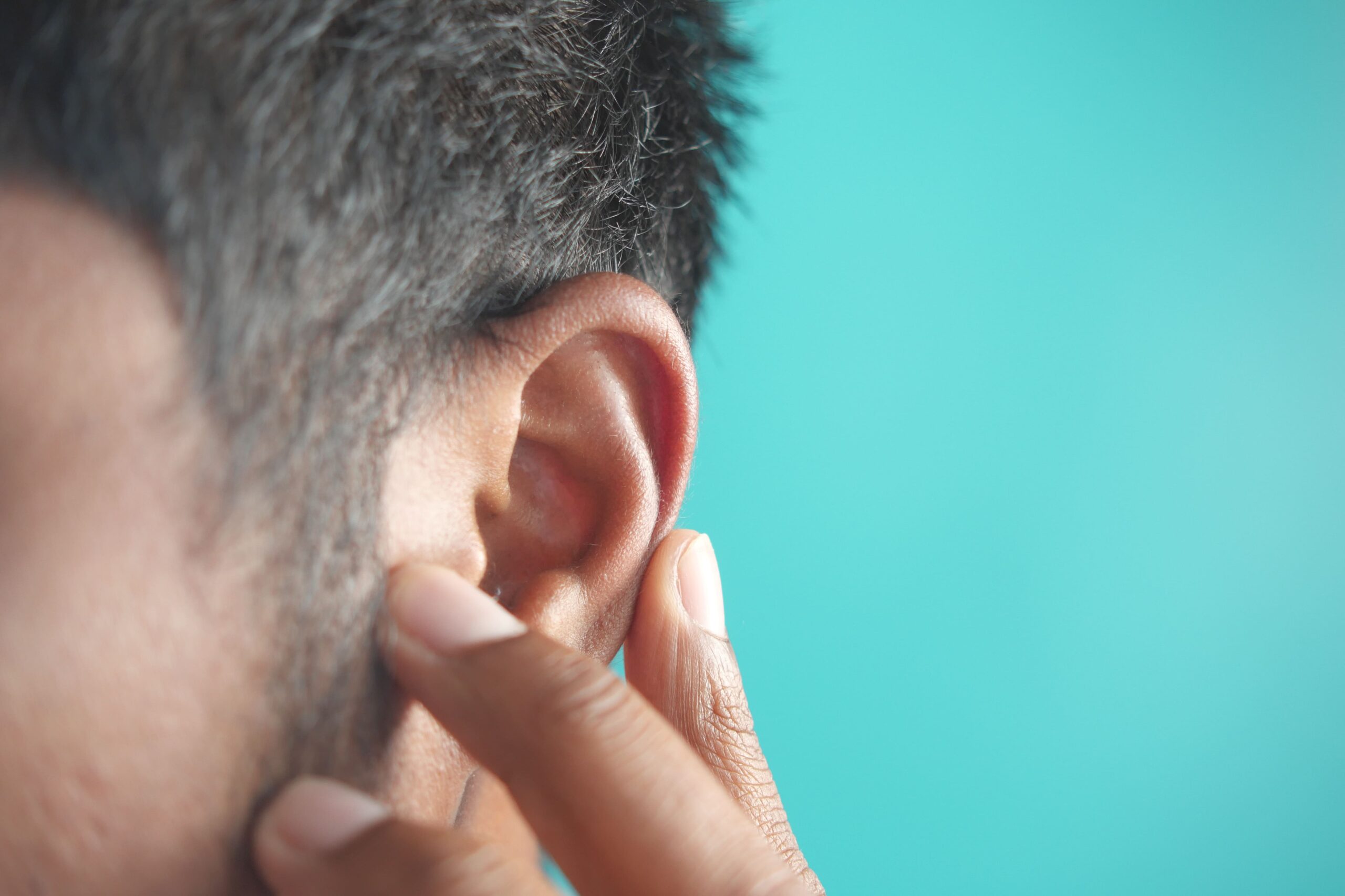
Signs & Symptoms
Early Symptoms
- Persistent foul-smelling ear discharge that doesn’t respond to antibiotics
- Gradual hearing loss in the affected ear
- Feeling of fullness or pressure in the ear
- Occasional mild ear pain or discomfort
- Debris visible in the ear canal during examination
Progressive Symptoms
- Increasing hearing loss is becoming more noticeable in daily activities
- Recurring ear infections despite treatment
- Persistent drainage that may be bloody
- Dizziness or mild balance problems
- Tinnitus (ringing or buzzing in the ear)
- Facial muscle weakness on the affected side
Advanced Symptoms
- Hearing loss or deafness in the affected ear
- Vertigo and balance difficulties
- Facial paralysis from facial nerve involvement
- Headaches suggesting intracranial involvement
- Confusion or altered mental state indicating brain complications
Cholesteatoma symptoms develop gradually. The hallmark sign is persistent, foul-smelling ear discharge that doesn’t improve with standard antibiotic treatment. As a cholesteatoma grows, it may erode surrounding structures, worsening symptoms.
Experiencing these symptoms?
Consult an ENT specialist for an accurate diagnosis and treatment plan.

When to See an ENT Specialist
Seek immediate medical attention if you experience sudden hearing loss, facial weakness, severe vertigo, or intense headaches with ear symptoms. These may indicate serious complications requiring urgent intervention. Schedule a consultation with an ENT specialist if you notice persistent ear discharge lasting more than two weeks, especially if it smells foul or contains blood. Progressive hearing loss, even if gradual, warrants professional evaluation as treatment can aim to preserve remaining hearing function.
During your first consultation, the ENT specialist typically conducts a thorough examination using an otoscope or microscope to visualise the ear canal and eardrum. They’ll review your medical history, focusing on previous ear infections, surgeries, or trauma. The specialist typically assesses your hearing and may identify characteristic signs of cholesteatoma, such as keratin debris, eardrum retraction pockets, or granulation tissue. This initial evaluation determines the extent of investigation needed and the urgency of treatment.
Early intervention is essential for cholesteatoma, as the growth continues to expand, causing progressive damage. Timely treatment may help preserve hearing, reduce the risk of facial nerve damage, and avoid serious complications such as meningitis or brain abscess.
Diagnosis & Testing Methods
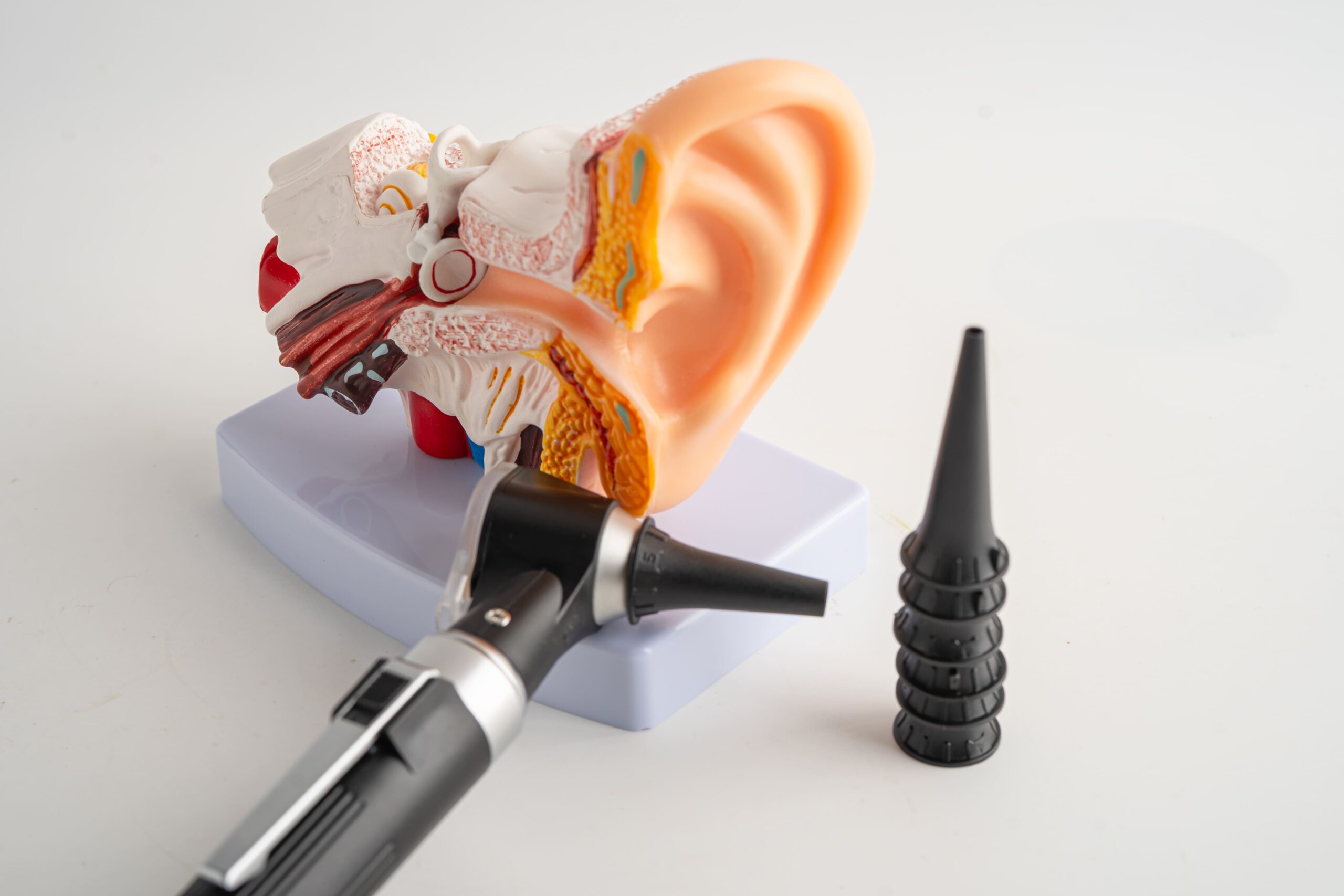
Accurate diagnosis of cholesteatoma requires examination and imaging studies to determine the extent of the disease and plan appropriate treatment. Your ENT specialist may begin with otomicroscopy, a microscopic examination of the ear. This allows visualisation of characteristic features such as white keratin debris, retraction pockets, or erosion of the ear canal bone.
Audiometry testing evaluates the degree and type of hearing loss. This test measures hearing sensitivity across different frequencies and determines whether hearing loss is conductive (from middle ear damage) or sensorineural (from inner ear involvement). Tympanometry assesses eardrum mobility and middle ear pressure, helping identify perforations or fluid accumulation.
High-resolution CT scanning is commonly used for imaging cholesteatoma. This scan can reveal the extent of bone erosion, involvement of critical structures such as the facial nerve or inner ear, and aid surgical planning. In some cases, MRI may be recommended to differentiate cholesteatoma from other conditions or detect residual disease after surgery.
Your ENT specialist may review all findings to determine the extent of the cholesteatoma and develop an individualised treatment strategy.
Treatment Options Overview
Medical Management
Cholesteatoma requires surgical treatment, but medical management provides important support. Antibiotic ear drops help control infection and reduce discharge before surgery. Your ENT specialist may prescribe fluoroquinolone drops, which penetrate well and cover common ear bacteria. Regular ear cleaning (aural toilet) in the clinic removes debris and allows better medication penetration. This involves gentle suction under microscopic guidance.
Canal Wall Up Mastoidectomy
This surgical technique removes the cholesteatoma whilst preserving the posterior ear canal wall. The surgeon accesses the middle ear and mastoid bone through an incision behind the ear, carefully removing cholesteatoma tissue whilst maintaining normal ear anatomy. This approach preserves the natural shape of the ear canal, avoiding long-term maintenance issues. This technique may be considered when the cholesteatoma is limited and complete removal appears achievable.
Canal Wall Down Mastoidectomy
For extensive cholesteatoma, canal wall down mastoidectomy provides better disease clearance and visualisation. The surgeon removes the posterior canal wall, creating a larger cavity that combines the ear canal with the mastoid bowl. This aims to enable complete cholesteatoma removal and may reduce recurrence risk, but requires lifelong periodic cleaning. The larger cavity may accumulate debris and be prone to water infiltration.
Endoscopic Ear Surgery
Endoscopic techniques use cameras and instruments inserted through the ear canal, avoiding external incisions. This approach provides visualisation of areas where cholesteatoma commonly recurs. Suitable for selected cases with limited disease, endoscopic surgery may offer faster recovery, less postoperative pain, and no visible scarring.
Ossicular Chain Reconstruction
When a cholesteatoma erodes the tiny ear bones (ossicles), causing hearing loss, reconstruction aims to restore the sound-conduction mechanism. This may involve repositioning remaining ossicles or placing prosthetic devices made of titanium or hydroxyapatite. Reconstruction may be performed during initial surgery or as a staged procedure, allowing the ear to heal first. The extent of hearing improvement varies depending on individual anatomy and disease extent.
Second-Look Surgery
Due to cholesteatoma’s tendency to recur, second-look surgery is often planned after initial treatment. This procedure checks for residual or recurrent disease in areas that are initially difficult to visualise. If cholesteatoma is found, it can be removed. If the ear is clear, ossicular reconstruction to improve hearing may be performed. Some centres now use diffusion-weighted MRI as an alternative to second-look surgery.
Every patient’s condition is unique.
Our ENT Specialist can assess your specific situation and recommend the most suitable treatment.
Complications if Left Untreated
Untreated cholesteatoma may progressively affect surrounding structures, potentially leading to serious complications. The expanding mass can erode through bone, creating pathways for infection to spread beyond the ear. Hearing loss may occur as the cholesteatoma affects the ossicles and damages the inner ear. Once the inner ear is affected, hearing loss may become difficult to reverse even with treatment.
Facial nerve weakness may develop when cholesteatoma erodes the bony canal protecting the facial nerve. This can cause weakness or paralysis of facial muscles on the affected side, affecting eye closure, smile, and facial expression. Labyrinthitis, an inner ear infection, can cause vertigo, nausea, and hearing loss. Patients may experience difficulty with walking or maintaining balance.
Intracranial complications, though uncommon, can be serious. Cholesteatoma may erode through the skull base, potentially causing meningitis, brain abscess, or sigmoid sinus thrombosis. These complications may present with headache, fever, confusion, and neurological symptoms. Quality of life may be affected as patients deal with chronic discharge and progressive functional impairment.
This information is provided for educational purposes and should not replace professional medical consultation.
Medisave & Insurance Shield Plan approved
Your ENT procedure may be eligible for Medisave claims, with the claimable amount varying based on the procedure’s complexity. For additional options, including the use of your insurance or Integrated Shield Plan, reach out to our friendly clinic staff today for assistance.


Frequently Asked Questions (FAQ)
Is cholesteatoma surgery painful, and what is the recovery like?
Cholesteatoma surgery is performed under general anaesthesia, so you won’t feel anything during the procedure. Post-operative discomfort is generally mild to moderate and well-controlled with pain medication. Many patients describe it as pressure or fullness rather than sharp pain. You’ll have a bandage around your head for the first day, which is removed before discharge. Recovery varies depending on the surgical technique used, but patients may return to desk work within a timeframe determined by their surgeon. Complete healing may take several weeks, during which you’ll need to keep the ear dry and attend follow-up appointments for ear cleaning.
Can cholesteatoma come back after surgery?
Recurrence is possible, with rates varying depending on various factors. The extent of initial disease, the surgical technique used, and eustachian tube function all influence the risk of recurrence. Regular follow-up is crucial, with frequency determined by your ENT specialist. Your ENT specialist may recommend second-look surgery or MRI scanning to detect early recurrence when it may be easier to treat.
Will I regain my hearing after cholesteatoma surgery?
Hearing outcomes depend on the extent of damage before surgery. If the cholesteatoma has affected the eardrum and ossicles, reconstruction may help improve hearing. However, if the inner ear is damaged, hearing improvement may be limited. Your pre-operative hearing test helps predict potential outcomes. Some patients may benefit from hearing aids after surgery. The primary goal of surgery is to remove disease and prevent complications, with hearing improvement as a secondary objective when safely achievable.
How long will I need to take off work or school?
Recovery time varies by individual and depends on the type of work or activities involved. Those with desk jobs may return sooner, while jobs requiring physical activity, exposure to dust, or water contact may require a longer recovery. Children typically return to school based on their surgeon’s recommendations, but should avoid physical education and swimming for a period determined by their healthcare team. Your ENT specialist can provide specific recommendations based on your surgery type and occupation. Plan for several follow-up appointments during recovery for ear cleaning and monitoring.
Can cholesteatoma be treated without surgery?
Cholesteatoma cannot be treated definitively without surgery. Unlike simple ear infections that respond to antibiotics, cholesteatoma is a physical mass of skin cells that must be surgically removed. Medical treatment with ear drops and cleaning may temporarily control symptoms, but cannot stop progression. Delaying surgery allows the cholesteatoma to grow larger, potentially causing more damage and making eventual surgery more complex. The primary treatment approach is complete surgical excision.
What are the long-term follow-up requirements after cholesteatoma surgery?
Long-term follow-up is essential after cholesteatoma surgery to monitor for recurrence and maintain ear health. Initially, you’ll need appointments for ear cleaning as it heals. Once healed, follow-up continues based on your surgeon’s recommendations. Patients who have undergone canal wall-down surgery may require periodic cleaning. These appointments include ear examination and may involve hearing tests or imaging. Consistent follow-up allows early detection and treatment of any recurrence. Follow-up schedules and requirements may vary by individual case.
Conclusion
Cholesteatoma is a serious ear condition that requires surgical treatment to help prevent potentially serious complications. While the diagnosis may be concerning, contemporary surgical techniques can offer good outcomes when the condition is addressed promptly. The approach to treatment involves early detection, surgical removal, and ongoing follow-up care. With appropriate treatment, patients may avoid serious complications, and some may experience hearing improvement. Whether you’re experiencing persistent ear discharge, hearing loss, or have been diagnosed with cholesteatoma, seeking care from an ENT specialist in Singapore can help you receive appropriate treatment tailored to your specific situation.
Take the First Step Towards Better Ear Health
Living with cholesteatoma symptoms can be concerning, but you don’t have to face it alone. Our ENT Specialist has experience diagnosing and treating cholesteatoma using contemporary evidence-based surgical techniques.

Dr Gan Eng Cern
MBBS
MRCS (Edin)
mmed (orl)
FAMS
With over 15 years of experience, Dr Gan specialises in the comprehensive management of a broad range of conditions related to the ear, nose and throat (ENT), and head and neck.
Dr. Gan has contributed to the academic field as a Senior Clinical Lecturer at the Yong Loo Lin School of Medicine, National University of Singapore. He underwent his subspecialty training in Rhinology (Nose & Sinus conditions) and Endoscopic Skull Base Surgery at the renowned St Paul’s Sinus Center, part of the University of British Columbia in Vancouver, Canada. He is recognised for his extensive research work, with numerous contributions to reputable international ENT journals. Dr Gan is also highly sought after as a speaker and has shared his surgical knowledge as a surgical dissection teacher at various prominent ENT conferences and courses.
- 2020 – Reader’s Choice Gold Award for Best ENT Specialist (Expat Living Singapore)
- 2016 – Best Educator Award (Eastern Health Alliance)
- 2016 – “Wow” Award (Patient Compliment)
- 2014 – Eastern Health Alliance Caring Award – Silver
- 2014 – 19th Yahya Cohen Memorial Lectureship (awarded by the College of Surgeons, Academy of Medicine Singapore for best scientific surgical paper)
- 2012 – Human Manpower Development Award (Ministry of Health, Singapore)
- 2007 – Singhealth Best Doctor Award
Qualifications
- MBBS – Bachelor of Science, Bachelor of Medicine, Bachelor of Surgery (University of New South Wales, Sydney, Australia)
- MRCS (Edin) – Member of the Royal College of Surgeons Edinburgh, United Kingdom
- MMed (ORL) – Master of Medicine in ENT (National University of Singapore)
- FAMS – Fellow of the Academy of Medicine Singapore

Make an Enquiry
Fill up the form and our friendly clinic staff will get back to you promptly.
Our Location
A Specialist Clinic for Sinus, Snoring & ENT
38 Irrawaddy Road #08-45
Mount Elizabeth Novena Hospital
Singapore 329563
Mon – Fri: 9:00 am – 5:00 pm
Sat: 8:30 am – 12:00 pm