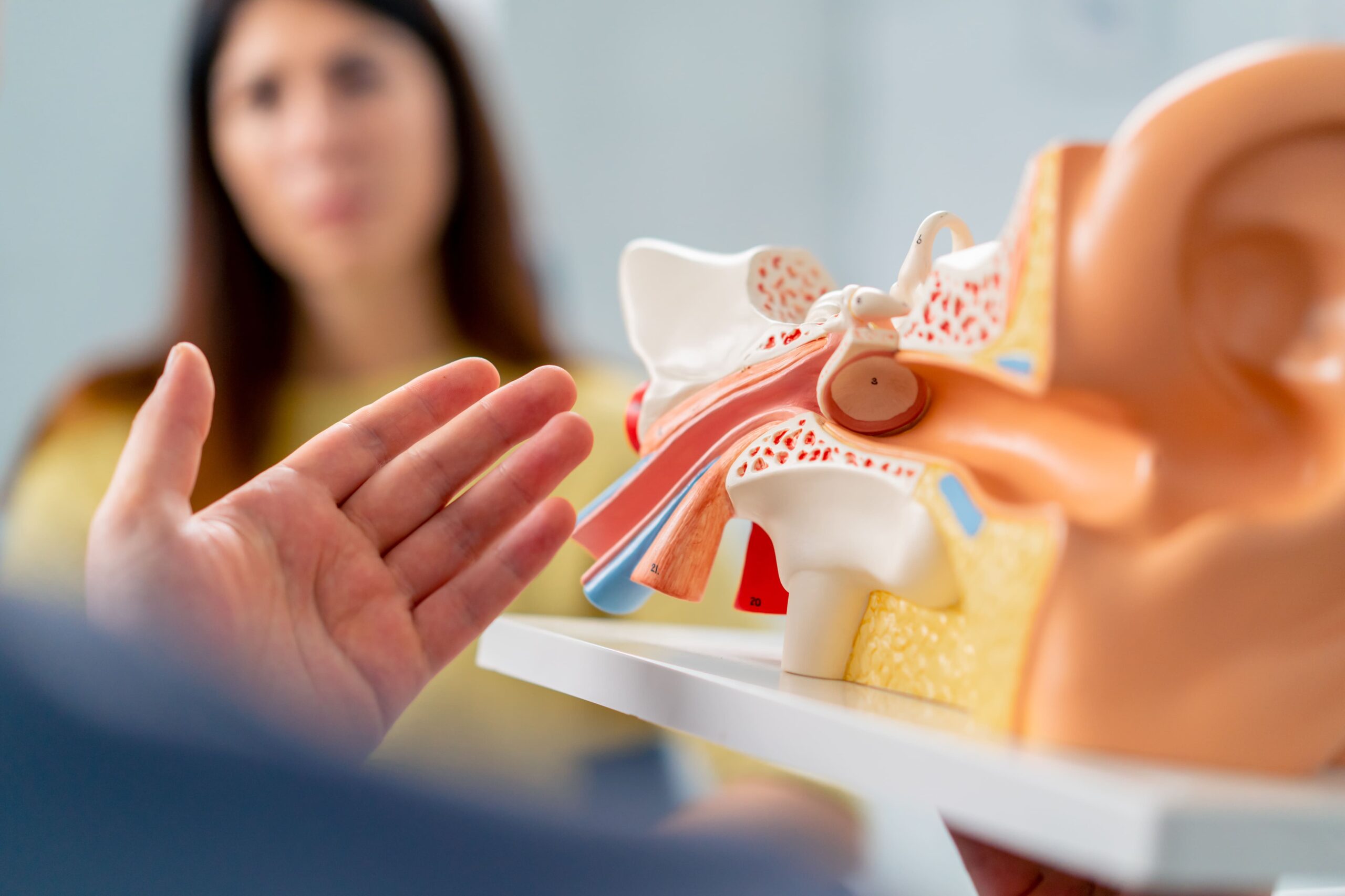
Accurate diagnosis begins with otoscopic examination, where an ENT specialist uses a specialised instrument to visualise the eardrum directly. This examination reveals the perforation’s size, location, and condition of the surrounding tissue. Video otoscopy allows patients to see their own eardrum on a screen, enhancing understanding of their condition.
Audiometry testing measures hearing function across different frequencies and can help assess conductive hearing loss that may be associated with the perforation. This assessment can be valuable for monitoring treatment progress and determining surgical candidacy.
Tympanometry evaluates middle ear function by measuring eardrum mobility in response to pressure changes. A perforated eardrum may produce characteristic flat tympanogram patterns, which can help confirm the diagnosis.
CT imaging may be recommended for traumatic perforations or when complications are suspected. This detailed imaging can reveal temporal bone fractures, ossicular chain disruption, or cholesteatoma formation that might influence treatment planning.
Bacterial culture of ear discharge, when present, can help identify causative organisms and guide antibiotic selection for infection management before definitive treatment.