Fraud Blocker

Fluid in Ear Treatment in Singapore

Experiencing muffled hearing, ear fullness, or discomfort from fluid trapped in your ear can impact your daily life and may lead to complications if left untreated. Fluid in the ear, medically known as otitis media with effusion (OME), occurs when fluid accumulates behind the eardrum without signs of acute infection. This condition affects both children and adults in Singapore, and is commonly seen in young children due to their developing eustachian tubes. ENT specialists can provide evaluation and treatment options that aim to restore ear health and hearing clarity, using established medical approaches tailored to each patient’s specific condition. Individual results and treatment outcomes may vary.

doctor img
Dr Gan Eng Cern

MBBS

MRCS (Edin)

mmed (orl)

FAMS

Image fluid in ear Image fluid in ear

What is Fluid in Ear (Otitis Media with Effusion)?

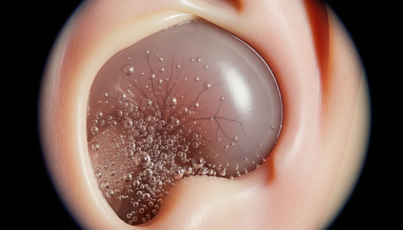
Fluid in the ear, or otitis media with effusion (OME), occurs when fluid accumulates in the middle ear space behind the eardrum without causing an active infection. This fluid buildup happens when the eustachian tube, which connects the middle ear to the back of the throat, becomes blocked or fails to function properly. The trapped fluid can be thin and watery or thick and glue-like, leading some to call this condition “glue ear.”

The condition differs from acute otitis media (middle ear infection) as it typically doesn’t cause severe pain or fever. The accumulated fluid can cause hearing difficulties, ear pressure, and balance problems. In Singapore’s humid climate, allergies, upper respiratory infections, and sinus problems can contribute to this condition. The fluid dampens sound vibrations, resulting in conductive hearing loss that can affect speech development in children and communication in adults.

Whilst fluid in the ear often resolves on its own, persistent cases may require medical intervention to help prevent complications such as chronic hearing loss, speech delays in children, or structural damage to the ear. Treatment approaches range from watchful waiting and medical management to surgical interventions, depending on the duration and severity of symptoms.

Who is a Suitable Candidate for Fluid in Ear Treatment?

Ideal Candidates

  • Children with persistent fluid lasting more than three months despite conservative management
  • Adults experiencing chronic ear fullness and hearing difficulties affecting work or daily activities
  • Patients with recurrent episodes of fluid accumulation following upper respiratory infections
  • Individuals with documented hearing loss on audiometry testing due to middle ear fluid
  • Children showing speech or language developmental delays associated with hearing impairment
  • Patients with structural abnormalities of the eustachian tube or palate requiring intervention
  • Those experiencing complications such as retraction pockets or adhesive otitis media
  • Individuals with fluid in ear accompanied by chronic sinusitis or allergic rhinitis

Contraindications

  • Active middle ear infection requiring antibiotic treatment before procedural intervention
  • Bleeding disorders that may increase surgical risks for procedures like myringotomy
  • Severe immunodeficiency where surgical intervention may pose increased infection risk
  • Anatomical abnormalities that make standard treatment approaches unsuitable
  • Uncontrolled allergies or chronic conditions that need stabilisation first

Suitable treatment requires thorough evaluation by an ENT specialist. Factors including symptom duration, degree of hearing loss, impact on quality of life, and presence of complications guide treatment decisions. In children, developmental considerations and the potential risk of long-term effects on speech and learning play important roles in treatment planning.

Treatment Techniques & Approaches

Medical Management

Medical management serves as the first-line treatment for fluid in ear, particularly in cases lasting less than three months. This approach includes medications to reduce inflammation, manage allergies, and improve eustachian tube function. Nasal corticosteroid sprays help reduce inflammation in the nasal passages and eustachian tube opening. Antihistamines and decongestants may provide relief when allergies contribute to the condition. Auto-insufflation techniques, where patients gently blow air into their middle ear through specialised devices, can help open the eustachian tube and promote fluid drainage.

Myringotomy with Ventilation Tube Insertion

Myringotomy with ventilation tube insertion is a surgical treatment option for persistent fluid in ear. During this procedure, the ENT surgeon creates a small incision in the eardrum (myringotomy) and places a tiny ventilation tube (grommet) to allow continuous drainage and ventilation of the middle ear. The tubes typically remain in place before naturally extruding. This procedure aims to bypass the dysfunctional eustachian tube, and may help improve hearing and reduce fluid reaccumulation.

Balloon Eustachian Tuboplasty

Balloon eustachian tuboplasty represents a treatment option for adults with chronic eustachian tube dysfunction. This minimally invasive procedure involves inserting a small balloon catheter through the nose into the eustachian tube opening. The balloon is inflated to dilate the tube, which may improve its function and reduce fluid accumulation. This technique addresses the underlying cause rather than just managing symptoms.

Technology & Equipment Used

Modern otoscopy and endoscopic equipment allow precise visualisation of the ear structures and fluid characteristics. Tympanometry provides objective measurement of middle ear function and fluid presence. Operating microscopes enable accurate surgical intervention during myringotomy and tube placement. Balloon dilation systems offer controlled, measured dilation of the eustachian tube in appropriate candidates.

Wondering which approach is right for you?

Our ENT Specialist will evaluate your specific needs and recommend the most suitable technique.

Human head sinus structure

The Treatment Process

Pre-Treatment Preparation

Before treatment begins, comprehensive evaluation includes detailed medical history, physical examination, and hearing assessment. Otoscopy allows direct visualisation of the eardrum and any visible fluid. Tympanometry measures middle ear pressure and mobility, confirming fluid presence. Audiometry testing determines the degree of hearing loss. For children, age-appropriate hearing tests ensure accurate assessment.

Patients scheduled for surgical intervention receive specific pre-operative instructions. These include fasting requirements for procedures under general anaesthesia, typically nothing by mouth after midnight. Medications that affect blood clotting may need temporary discontinuation. Parents of paediatric patients receive detailed information about the procedure and post-operative care requirements.

During the Procedure

For medical management, treatment involves regular medication administration and follow-up monitoring. Nasal spray techniques are demonstrated to ensure proper delivery to the eustachian tube area. Auto-insufflation devices require proper instruction for effective use at home.

Myringotomy with tube insertion is typically performed. Under general anaesthesia for children or local anaesthesia for adults, the surgeon uses an operating microscope to visualise the eardrum. A small incision is made, fluid is suctioned out, and a ventilation tube is carefully placed. The procedure may be performed on one or both ears as needed.

Balloon eustachian tuboplasty, performed under local or general anaesthesia, involves endoscopic guidance to position the balloon catheter. The balloon is inflated at controlled pressure, then deflated and removed.

Immediate Post-Treatment

Following medical management initiation, patients are monitored for medication response and side effects. Improvement in symptoms may take several weeks to become apparent. Regular follow-up ensures treatment effectiveness.

After myringotomy with tube insertion, patients recover from anaesthesia. Hearing improvement may be noticeable once anaesthesia effects resolve. Mild ear drainage may occur initially as remaining fluid exits through the tube. Pain is typically minimal and may be managed with simple analgesics. Patients typically return home within hours of the procedure.

Following balloon tuboplasty, patients may experience temporary ear fullness or mild discomfort. These sensations typically resolve. Hearing improvement may be gradual as eustachian tube function improves and any remaining fluid drains naturally.

Recovery & Aftercare

First 24-48 Hours

After ventilation tube insertion, keep ears dry during bathing to prevent water entering through the tubes. Ear plugs or cotton balls with petroleum jelly provide provide protection. Take any prescribed ear drops as directed. Mild drainage from the ears is normal and should be gently cleaned with a soft cloth. Pain medication is rarely needed beyond the first day.

Following balloon tuboplasty, patients may experience crackling or popping sounds as the eustachian tube begins functioning normally. Gentle nose blowing is encouraged to promote tube opening. Normal activities can resume immediately, though strenuous exercise should wait 24 hours.

First Week

Patients with ventilation tubes attend a follow-up appointment to ensure proper tube position and function. Hearing may show improvement by this time. Swimming precautions vary by surgeon preference; some allow surface swimming without ear protection whilst others recommend ear plugs for all water activities.

For medical management patients, consistent medication use and monitoring for improvement continues. Nasal spray technique is reinforced to ensure delivery. Auto-insufflation exercises are performed multiple times daily as prescribed.

Long-term Recovery

Ventilation tubes require regular monitoring to check positioning and ensure continued function. The tubes may extrude naturally, with the eardrum healing spontaneously. Some patients may require tube replacement if fluid recurs after extrusion.

Balloon tuboplasty patients may experience gradual improvement over several weeks to months as the eustachian tube maintains improved function. Regular follow-up with audiometry can confirm hearing improvement. Additional treatments may be considered if symptoms recur.

Medical management often continues for several months with gradual tapering based on response. Addressing underlying causes such as allergies or chronic sinusitis may help prevent recurrence. Environmental modifications, including allergen reduction and smoking cessation, can support long-term outcomes.

Our ENT Specialist provides comprehensive post-procedure support to help ensure recovery.

Schedule your consultation to learn more about what to expect.

Human head sinus structure

Benefits of Fluid in Ear Treatment

Treatment of fluid in ear may help restore hearing function for patients of all ages. Improvement in hearing ability can support better communication in various environments, though individual outcomes vary.

For children, timely treatment aims to prevent potential speech and language delays that may be associated with hearing difficulties during developmental periods. Academic participation might improve as children may better hear in classroom settings. Behavioural concerns related to hearing difficulties could potentially resolve with appropriate treatment.

Adults may experience relief from sensations of ear fullness and pressure. Balance concerns and dizziness associated with middle ear fluid might improve, though individual responses vary. Treatment can help reduce the risk of recurrent ear infections and potential complications.

Treatment may also provide psychological benefits by addressing concerns about hearing function and its impact on daily activities. Parents of affected children might experience reduced stress as their child’s hearing improves. Individual results and timelines may vary. This treatment is administered as part of a comprehensive plan supervised by a healthcare professional.

Risks & Potential Complications

Common Side Effects

Ventilation tube insertion may cause temporary ear drainage as residual fluid exits the middle ear. This drainage typically resolves within a few days but occasionally persists, requiring antibiotic ear drops. Tubes may become blocked with wax or debris, requiring cleaning during follow-up visits. Some patients experience increased sensitivity to loud sounds initially as hearing improves.

Medical management side effects depend on specific medications used. Nasal corticosteroids may cause temporary nasal irritation or mild nosebleeds. Antihistamines can cause drowsiness in some individuals. Decongestants may cause temporary sleep disturbance or increased heart rate in sensitive patients.

Rare Complications

Persistent eardrum perforation can occur after tube extrusion, potentially requiring surgical repair (tympanoplasty). Chronic ear drainage despite appropriate treatment may affect some patients and could indicate underlying middle ear disease. Premature tube extrusion or displacement into the middle ear occurs rarely but may necessitate removal or replacement.

Scarring of the eardrum (tympanosclerosis) can develop but rarely affects hearing significantly. Middle ear infection around the tube requires antibiotic treatment and occasionally tube removal. Cholesteatoma formation, though extremely rare with modern ventilation tubes, requires surgical intervention if it occurs.

ENT specialists aim to minimise these risks through careful patient selection, meticulous surgical technique, and comprehensive post-operative care. Regular monitoring allows early detection and management of any complications that may arise.

Cost Considerations

The cost of fluid in ear treatment varies significantly based on the chosen treatment approach, whether one or both ears require treatment, and the complexity of the individual case. Medical management costs include consultation fees, diagnostic tests such as audiometry and tympanometry, and ongoing medication expenses. These conservative treatments generally represent an economical option but may require extended treatment periods.

Surgical interventions such as myringotomy with tube insertion include surgeon fees, facility charges, anaesthesia costs, and the ventilation tubes themselves. Balloon eustachian tuboplasty, being a contemporary procedure, may involve different cost structures. Post-operative care, including follow-up visits and any necessary medications, contributes to overall treatment expenses.

The consultation with an ENT specialist includes comprehensive evaluation and treatment recommendations with associated cost estimates. Factors such as the need for general anaesthesia versus local anaesthesia, treatment of one or both ears, and any concurrent procedures affect the final cost. Quality care requires appropriate investment, and transparent pricing discussions during consultation can help patients make informed decisions about their treatment.

For Singaporeans & Singapore Permanent Residents

Medisave & Insurance Shield Plan approved

Your ENT procedure may be eligible for Medisave claims, with the claimable amount varying based on the procedure’s complexity. For additional options, including the use of your insurance or Integrated Shield Plan, reach out to our friendly clinic staff today for assistance.

Frequently Asked Questions (FAQ)

How long does fluid typically remain in the ear without treatment?

Fluid in the ear often resolves spontaneously within three months in many cases. However, some patients experience persistent fluid for six months or longer without intervention. Children tend to have longer duration due to their horizontal eustachian tubes and increased susceptibility to upper respiratory infections. The watchful waiting approach with regular monitoring is appropriate for the first three months unless hearing loss is significant or complications develop. After three months, treatment intervention becomes increasingly important to prevent long-term consequences.

Can fluid in ear return after successful treatment?

Recurrence of fluid in ear can occur, particularly in children with ongoing risk factors. After ventilation tubes extrude naturally, some patients experience fluid reaccumulation requiring additional treatment. Addressing underlying causes such as allergies, chronic sinusitis, or exposure to cigarette smoke may help reduce recurrence risk. Regular monitoring allows early detection of recurrence. Some children may require multiple sets of tubes during their early years until eustachian tube function matures. Adults with chronic eustachian tube dysfunction may need long-term management strategies.

Is surgery always necessary for treating fluid in ear?

Surgery is not always necessary for treating fluid in ear. Many cases resolve with conservative management including watchful waiting, medical treatment, and auto-insufflation exercises. Surgery becomes indicated when fluid persists beyond three months with documented hearing loss, when complications develop, or when the condition significantly impacts quality of life. Children with speech delays or learning difficulties related to hearing loss may benefit from earlier surgical intervention. The decision for surgery is individualised based on duration of symptoms, degree of hearing loss, and response to conservative measures.

What happens to ventilation tubes over time?

Ventilation tubes are designed to be temporary and typically remain in place for several months before naturally extruding (falling out) as the eardrum grows. The extrusion process is usually painless, and patients may notice the tube in ear wax or it may fall out unnoticed. After extrusion, the eardrum heals spontaneously in most cases within a few weeks. Regular monitoring ensures tubes remain properly positioned and functional while in place. Tubes that don’t extrude after an extended period may require surgical removal to prevent complications.

Can adults develop fluid in ear, or is it only a childhood condition?

While fluid in ear is more common in children, adults can definitely develop this condition. Adult cases often result from eustachian tube dysfunction caused by allergies, sinusitis, air pressure changes, or upper respiratory infections. Adults may also develop fluid in ear following radiation therapy to the head and neck or due to nasopharyngeal masses. The treatment approach in adults may differ, with options like balloon eustachian tuboplasty available that aren’t typically used in children. Adult symptoms often focus more on ear pressure and hearing difficulty rather than developmental concerns.

How can I prevent fluid from accumulating in the ears?

Prevention strategies focus on maintaining healthy eustachian tube function and reducing risk factors. Managing allergies with appropriate medications, treating sinus infections promptly, and avoiding cigarette smoke exposure may help prevent fluid accumulation. For children, breastfeeding during infancy and avoiding bottle feeding while lying flat may reduce risk. During upper respiratory infections, gentle nose blowing and avoiding forceful sniffing help maintain eustachian tube patency. Regular follow-up with an ENT specialist for those with recurrent problems allows early intervention before significant fluid accumulates.

Conclusion

Fluid in ear, while common, requires proper evaluation and treatment to prevent complications and restore quality of life. Whether affecting children’s development or adults’ daily functioning, this condition may respond to various treatment approaches when appropriately selected and implemented. From conservative medical management to surgical interventions like ventilation tube insertion or balloon tuboplasty, modern ENT care offers options tailored to individual needs. Timely evaluation and accurate diagnosis support better outcomes and help guide the most suitable intervention based on symptom duration, severity, and patient-specific factors. Consulting an ENT specialist in Singapore can support effective management and long-term ear function improvement.

Ready to Take the Next Step?

If you’re experiencing persistent ear fullness, hearing difficulties, or your child shows signs of fluid in ear, our ENT Specialist can help you understand if treatment may be suitable for your needs. With experience in managing fluid in ear conditions, we provide personalised care throughout your journey.

Human head sinus structure
ENT Specialist in Singapore | Dr Gan Eng Cern

Dr Gan Eng Cern

MBBS

MRCS (Edin)

mmed (orl)

FAMS

With over 15 years of experience, Dr Gan specialises in the comprehensive management of a broad range of conditions related to the ear, nose and throat (ENT), and head and neck.

Dr. Gan has contributed to the academic field as a Senior Clinical Lecturer at the Yong Loo Lin School of Medicine, National University of Singapore. He underwent his subspecialty training in Rhinology (Nose & Sinus conditions) and Endoscopic Skull Base Surgery at the renowned St Paul’s Sinus Center, part of the University of British Columbia in Vancouver, Canada. He is recognised for his extensive research work, with numerous contributions to reputable international ENT journals. Dr Gan is also highly sought after as a speaker and has shared his surgical knowledge as a surgical dissection teacher at various prominent ENT conferences and courses.

  • 2020 – Reader’s Choice Gold Award for Best ENT Specialist (Expat Living Singapore)
  • 2016 – Best Educator Award (Eastern Health Alliance)
  • 2016 – “Wow” Award (Patient Compliment)
  • 2014 – Eastern Health Alliance Caring Award – Silver
  • 2014 – 19th Yahya Cohen Memorial Lectureship (awarded by the College of Surgeons, Academy of Medicine Singapore for best scientific surgical paper)
  • 2012 – Human Manpower Development Award (Ministry of Health, Singapore)
  • 2007 – Singhealth Best Doctor Award

Qualifications

  • MBBS – Bachelor of Science, Bachelor of Medicine, Bachelor of Surgery (University of New South Wales, Sydney, Australia)
  • MRCS (Edin) – Member of the Royal College of Surgeons Edinburgh, United Kingdom
  • MMed (ORL) – Master of Medicine in ENT (National University of Singapore)
  • FAMS – Fellow of the Academy of Medicine Singapore
Singapore ENT specialist performing a procedure

Make an Enquiry

Fill up the form and our friendly clinic staff will get back to you promptly.

    Our Location

    A Specialist Clinic for Sinus, Snoring & ENT

    38 Irrawaddy Road #08-45
    Mount Elizabeth Novena Hospital
    Singapore 329563

    Mon – Fri: 9:00 am – 5:00 pm
    Sat: 8:30 am – 12:00 pm